
Master Thesis "A brief introduction into the “eyes” of cyanobacteria (CBCRs)"
Biophysical study to understand the green/blue light reception of a microorganism.
Abstract:
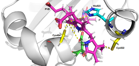
The aim of this study was to elucidate the time constants of the conformational changes of the chromophore phycoviolobilin (PVB) incorporated in the green-blue photoreceptor TePixJ, from the cyanobacterium Thermosynechococcus elongatus BP-1, upon illumination. The photoreceptor protein was expressed in E. coli and purified via nickel affinity chromatography as well as ion exchange chromatography. The photoconversion mechanism was analysed by flash photolysis experiments, performed along different pH-values. These experiments revealed that the Pg-state to Pb-state and the back conversion mechanism are, counterintuitively, fast at high pH-values but slows down at low pH-values. These effects are probably caused by the protonation state of a nearby histidine(523). However, the steady state UV/Vis measurements showed no alteration in absorption along the different pH-values. To investigate the binding properties of the chromophore in the Pb state, a steady state infrared spectra (FTIR) was recorded, revealing the formation of the second C-S bond between the chromophore and the cysteine(494).
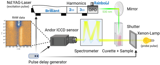
Time-resolved UV/Vis spectroscopy: is a technique used to study the kinetics of chemical reactions involving light-sensitive species. This technique has been widely used in the fields of photochemistry, biology to study a wide range of phenomena, including photodegradation, and photoinduced electron transfer. Time-resolved UV/Vis spectroscopy can also be used to determine the rate constants for the individual steps in the photoreceptors reaction cycle, such as the rate of electron transfer or the rate of protonation/deprotonation. This information can be used to construct detailed models of the photoreceptors function, which can aid in the design of new photoreceptor-based optogenetic tools. Here we measured transient UV/vis absorption spectra to elucidate the photocycle mechanism of phycocyanobilin incorporated into TePixJ.
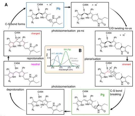
Photocycle of the light sensing protein:
The structures of TePixJ in both states (Pg and Pb) have been studied through X-ray crystallography and solution phase NMR. These studies suggest that photoconversion from Pb → Pg involves an initial photoisomerization of the bilin cofactor, which then triggers a series of structural changes in the protein. These changes include the a planarization and breakage and reformation of the C10-Cys494 thioether bond, opposite rotations of the A and D pyrrole rings, sliding of the bilin in the binding pocket, appearance of a disordered extended region including Cys494, and changes in the protein backbone. A current model of this mechanism can be seen on the right. To better understand those changes and the timing of this intricate mechanism, we used time-resolved UV/Vis spectroscopy.
The full Thesis you can download below.
